New paper in Nanoscale: “Differential labelling of human sub-cellular compartments with fluorescent dye esters and expansion microscopy”
Monday 13 November 2023
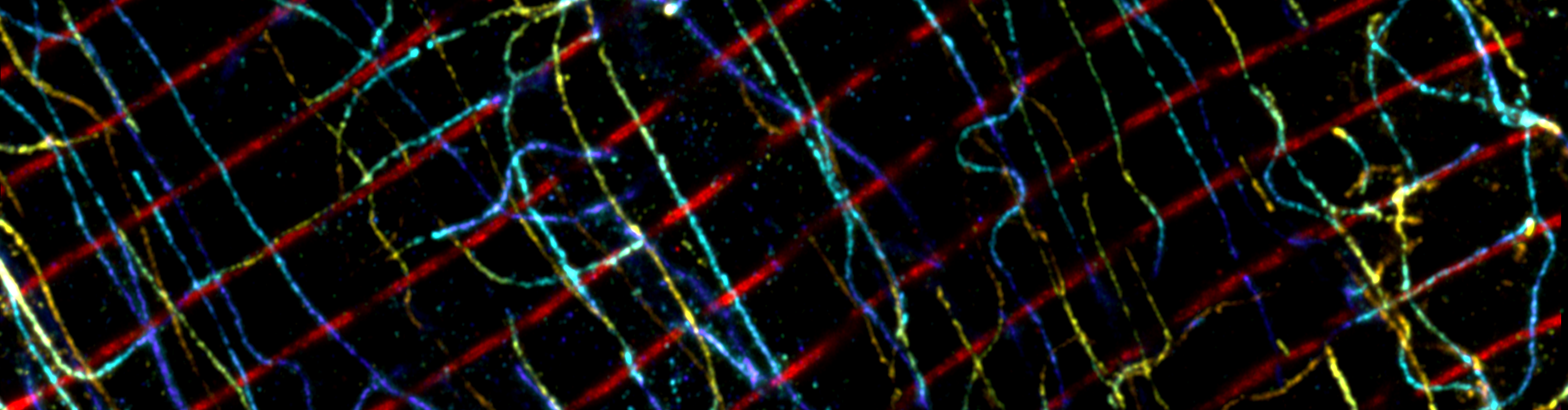
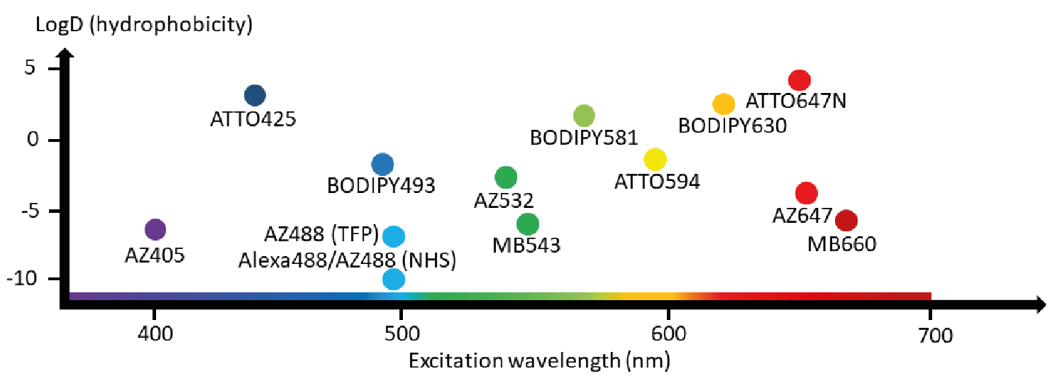
Our latest paper has been published in Nanoscale. We show how fluorescent esters can be used as versatile imaging probes capable of revealing an array of subcellular compartments, making use of human cell lines such as HeLa cells. These esters work by binding to free amine groups, effectively labelling the proteome in a non-descript way to reveal the overall cellular architecture. What makes these probes particularly versatile is that the labelling patterns differ depending on the properties of the conjugated dye molecule, allowing different dye esters to favour subtly different compartments depending on their lipophilicity. While we used expansion microscopy for improved resolution to see finer structures, these fluorescent ester imaging probes are very much useful for non-super-resolution imaging like confocal microscopy.
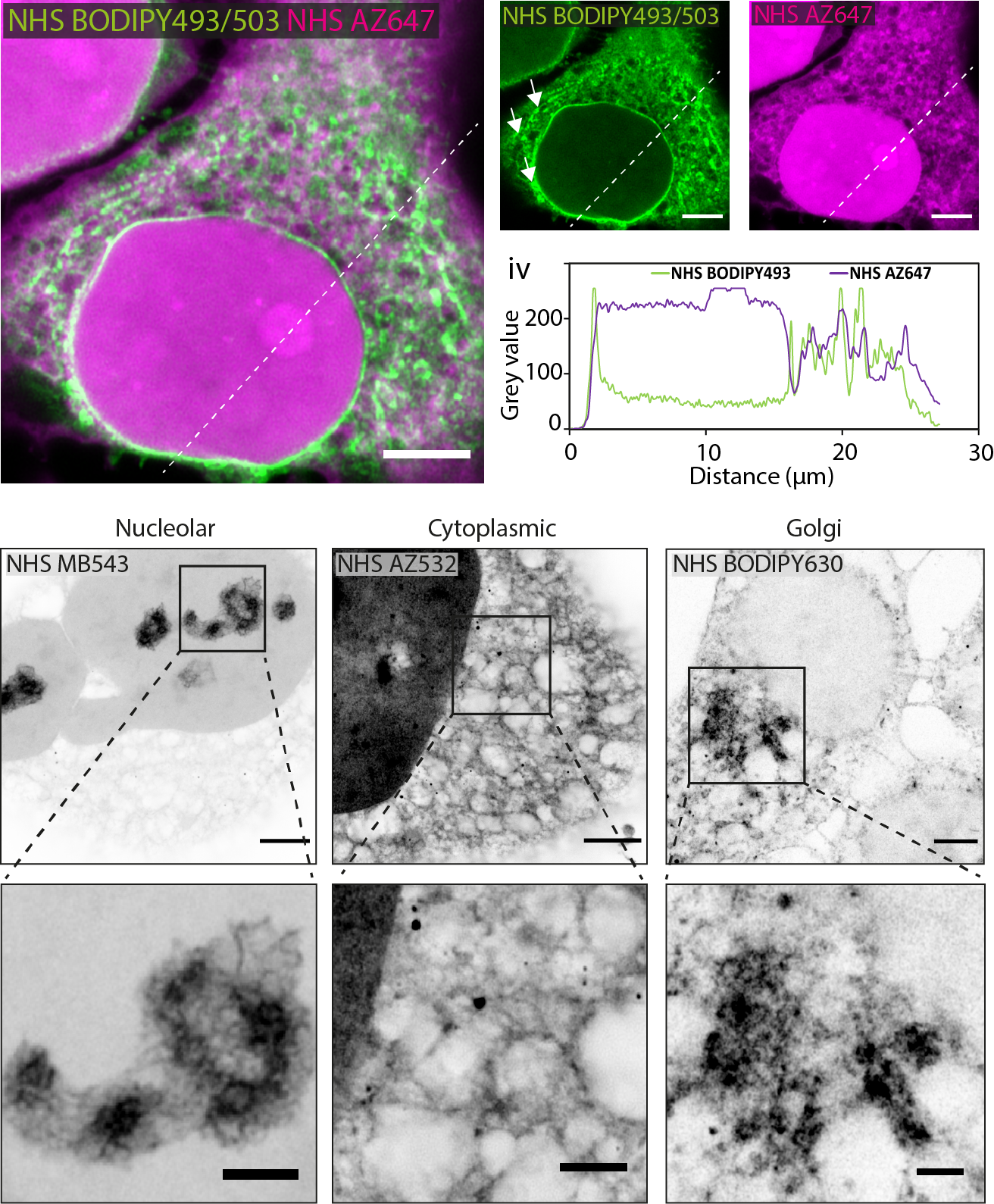
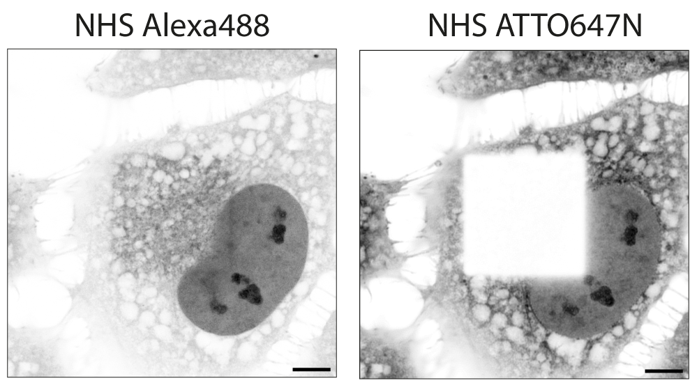
In the paper you can find catalogues of images for 14 esters conjugated to a bunch of dyes (Alexa, BODIPY and ATTO families), each possessing different charge and hydrophobic properties and spread across the visible spectrum.
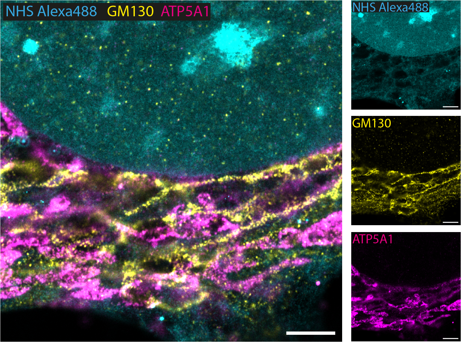
Esters applied by themselves are incredibly useful for visualising overall morphology, but they are perhaps most powerful when applied as a counterstain alongside specific labels. For instance, we show how esters can be used in tandem with antibody immunolabelling to give valuable contextual information, revealing each molecular target in relation to the broader cell architecture.
We also report new intricacies in labelling patterns which arise when multiple esters are added together through multi-ester interactions (involving FRET between the molecules), and which also depend on the timepoint of ester addition.
If you are a microscopist who studies cell biology I would strongly encourage you give these fluorescent esters a try, in our hands they have allowed extremely useful visualisations of morphology in a range of sample types, from cell monolayers to multi-cellular specimens. One example study in the model organism C. elegans, in collaboration with Dr Kin Man Suen from the University of Leeds, made use of esters to reveal membraneless compartments known as P granules, and allowed us to assess the relative arrangement of endogenously labelled proteins.
Many thanks to the co-authors Dr Izzy Jayasinghe, Dr Kin Man Suen, Tayla Shakespeare, Raj Seehra and Michael Spencer. Thanks too to Darren and Nick in the LMF imaging facility at the University of Sheffield, as well as the funders IBIN and UKRI who made this research possible!
The above is just a snippet of the research, read the new paper in Nanoscale in its entirety here.